Cardiovascular Function Following Prolonged Firefighting
Summary
Funding provided by: Department of Homeland Security, United States Fire Administration - Assistance to Firefighters Grant Program: Fire Prevention and Safety Grant
While sudden cardiac events are considered responsible for the highest percentage of line of duty deaths each year, the “cause” of these cardiovascular deaths is routinely attributed to “overexertion”. Yet, it is unclear how “overexertion” leads to sudden cardiac events and why some individuals are vulnerable to “overexertion” while others are not. Despite the discouraging national statistics relative to sudden cardiac events suffered by firefighters in the line of duty, and national efforts to reverse this situation, surprising little research had been conducted to investigate the nature of the “overexertion” that is reported to lead to sudden cardiac events in firefighters, or to systematically document the extent of the cardiovascular dysfunction caused by strenuous firefighting.
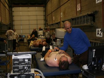
This study was designed to a) investigate the nature of overexertion related to firefighting, particularly as it applies to the cardiovascular system, b) examine cardiovascular characteristics (novel risk factors and early indicators of cardiovascular disease) of firefighters that may make them vulnerable to dangerous cardiovascular changes during firefighting, and c) explore the effectiveness of a nutritional supplement (Vitamin C) to mitigate detrimental cardiovascular responses. Firefighters were tested immediately before and after a 3-4 hour long period of intermittent live fire training exercises. Firefighting drills varied, but generally consisted of 4-5 evolutions lasting 15-25 minutes with 10-15 minutes of rest between each evolution. Each evolution included coordinated fireground operations between engine and truck companies in a variety of training environments.
Heart function was assessed through: a myocardial function assessment; a cardiac systolic and diastolic function test; Color Tissue Doppler imaging; and pulse contour analysis-derived indices of cardiac function. Vascular function was assessed through measurement of Augmentation Index (Pulse Wave Analysis), regional arterial stiffness (Pulse Wave Velocity), carotid artery compliance and β-stiffness index, and microvascular function (forearm resistance artery vasodilatory capacity). Blood samples were analyzed to assess hemostatic balance (via platelet number and function, coagulatory variables, and fibrinolytic factors).
